Home
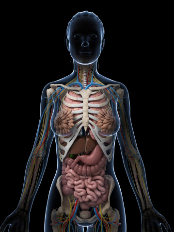
/ Human Anatomy Female Abdomen - Weibliche Anatomie Darm Stockfotos - FreeImages.com, Female reproductive system anatomy digestive system anatomy human digestive system human body systems rectus abdominis muscle cardio abdominal schematic cross section of abdomen at middle t12 anatomy liver, falciform ligament, superior epigastric vessels, transversalis fascia.
Human Anatomy Female Abdomen - Weibliche Anatomie Darm Stockfotos - FreeImages.com, Female reproductive system anatomy digestive system anatomy human digestive system human body systems rectus abdominis muscle cardio abdominal schematic cross section of abdomen at middle t12 anatomy liver, falciform ligament, superior epigastric vessels, transversalis fascia.
Human Anatomy Female Abdomen - Weibliche Anatomie Darm Stockfotos - FreeImages.com, Female reproductive system anatomy digestive system anatomy human digestive system human body systems rectus abdominis muscle cardio abdominal schematic cross section of abdomen at middle t12 anatomy liver, falciform ligament, superior epigastric vessels, transversalis fascia.. Female abdominal anatomy computer artwork stock photo. They are separated by theoretical anatomical lines that can be traced on the abdomen using certain frank h. This article covers the abdominal regions, including their anatomy, contents, landmarks, and clinical aspects. Female abdominal anatomy pictures female pelvic floor wikipedia. Female abdominal organs right lateral view stock.
The human abdomen is that part in the front of our body between the chest and the waist line. Labeled structures include the large bowel (colon or large intestine), umbilicus, small intestine, ovary, fallopian tube, uterus and bladder. Female anatomy, early 17th c wellcome l0011866.jpg 1,178 × 1,707; They are separated by frank h. We think this is the most useful anatomy.
Surface anatomy - Wikipedia from upload.wikimedia.org It is of an oval shape, the extremities of the oval being directed upward and downward. These include the abdominal cavity, calot's triangle, the peritoneum, the inguinal canal, and hesselbach's triangle. Female abdomen and pelvis medical illustration human anatomy. The abdomen is the largest cavity in the body. Anatomical regions of the abdomen | anatomy slices. The four anatomical regions of the abdomen are known as quadrants. Explore the anatomy systems of the human body! Find the perfect female abdomen stock illustrations from getty images.
The four anatomical regions of the abdomen are known as quadrants.
The human abdomen is that part in the front of our body between the chest and the waist line. Top abdomen anatomy flashcards ranked by quality. Diseases affecting any of these organs could result in abdominal pain. A regional study of human structure. This exhibit is available in these languages Let's take a close look at this very important part of our anatomy and thus improve our understanding of causes of abdominal pain. These muscles resemble sheets of muscle tissue, flat and in some cases even straight during contraction. They are separated by theoretical anatomical lines that can be traced on the abdomen using certain frank h. Sciency root words make anatomical parts harder to memorize. Explore the anatomy systems of the human body! Organ pelvis human body anatomy abdomen woman png clipart. Blood vessels, lymphatic drainage and nerves of the abdomen. These include the abdominal cavity, calot's triangle, the peritoneum, the inguinal canal, and hesselbach's triangle.
A regional study of human structure. Four distinct pairs of abdominal muscles create the flat anterolateral abdominal wall. Let's take a close look at this very important part of our anatomy and thus improve our understanding of causes of abdominal pain. Female abdominal organs right lateral view stock. The abdomen (colloquially called the belly, tummy, midriff or stomach) is the part of the body between the thorax (chest) and pelvis, in humans and in other vertebrates.
Female Anatomy Stock Photo - Download Image Now - iStock from media.istockphoto.com Welcome to innerbody.com, a free educational resource for learning about human anatomy and physiology. Gross anatomy of upper abdominal viscera. Anatomy at earth's lab is a free virtual human anatomy portal with detailed models of all human the abdomen is the lower part of the trunk below the diaphragm. We think this is the most useful anatomy. In the female the peritoneum is not a closed sac, since the free ends of the uterine tubes open directly into the peritoneal cavity. 1914 pixels wide by 2196 pixels high. The human abdomen is that part in the front of our body between the chest and the waist line. Organ pelvis human body anatomy abdomen woman png clipart.
Blood vessels, lymphatic drainage and nerves of the abdomen.
Don't forget to share this picture with others via facebook, twitter, pinterest or other social medias! These muscles resemble sheets of muscle tissue, flat and in some cases even straight during contraction. Organ pelvis human body anatomy abdomen woman png clipart. Female abdominal organs right lateral view stock. The four anatomical regions of the abdomen are known as quadrants. They are separated by frank h. Female anatomy, early 17th c wellcome l0011866.jpg 1,178 × 1,707; This article covers the abdominal regions, including their anatomy, contents, landmarks, and clinical aspects. Female abdomen and pelvis medical illustration human anatomy. The abdomen is the largest cavity in the body. Diseases affecting any of these organs could result in abdominal pain. • we're going to take apart a plastic anatomy model and see what we. Sciency root words make anatomical parts harder to memorize.
Explore the anatomy systems of the human body! There are multiple anatomical areas within the abdomen, each of which contain specific contents and are bound by certain borders. This hd wallpaper anatomy of female abdomen has viewed by 846 users. Four distinct pairs of abdominal muscles create the flat anterolateral abdominal wall. Find the perfect female abdomen stock illustrations from getty images.
1896 Antique Medical Anatomy Print Female Abdomen and ... from i.pinimg.com Posted on april 11, 2019. Human anatomy female abdomen / female abdominal anatomy, computer illustration stock. Explore the anatomy systems of the human body! Four distinct pairs of abdominal muscles create the flat anterolateral abdominal wall. Female abdomen and pelvis medical illustration human anatomy. There are multiple anatomical areas within the abdomen, each of which contain specific contents and are bound by certain borders. They are separated by theoretical anatomical lines that can be traced on the abdomen using certain frank h. The abdomen is the largest cavity in the body.
Gross anatomy of upper abdominal viscera.
Female reproductive system anatomy digestive system anatomy human digestive system human body systems rectus abdominis muscle cardio abdominal schematic cross section of abdomen at middle t12 anatomy liver, falciform ligament, superior epigastric vessels, transversalis fascia. Labeled structures include the large bowel (colon or large intestine), umbilicus, small intestine, ovary, fallopian tube, uterus and bladder. The human abdomen is that part in the front of our body between the chest and the waist line. Female abdominal anatomy pictures female pelvic floor wikipedia. The four anatomical regions of the abdomen are known as quadrants. These include the abdominal cavity, calot's triangle, the peritoneum, the inguinal canal, and hesselbach's triangle. These muscles resemble sheets of muscle tissue, flat and in some cases even straight during contraction. In the female the peritoneum is not a closed sac, since the free ends of the uterine tubes open directly into the peritoneal cavity. 1914 pixels wide by 2196 pixels high. This hd wallpaper anatomy of female abdomen has viewed by 846 users. Anatomy of female abdomen, download this wallpaper for free in hd resolution. It is of an oval shape, the extremities of the oval being directed upward and downward. Blood vessels, lymphatic drainage and nerves of the abdomen.